
Fig. S7
- ID
- ZDB-FIG-180828-50
- Publication
- Phan et al., 2018 - Neutrophils use superoxide to control bacterial infection at a distance
- Other Figures
- All Figure Page
- Back to All Figure Page
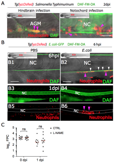
Recruited neutrophils do not produce detectable Nitric Oxide. (A) Nitric oxide is produced by neutrophils in the AGM following Salmonella Typhimurium infection. Two dpf tg(lyz:DsRed) embryos were infected in the hindbrain or in the notochord with Salmonella Typhimurium. At 2 dpi Nitric oxide was detected with DAF-FM-DA (green) using confocal microscopy. Representative overlay of maximum projections of multi scan acquisitions (DsRed and DAF-FM-DA) with transmitted light images shows that Nitric oxide is produced by neutrophils in the AGM (A left panel) and in the notochord (A right panel), but not in the recruited neutrophils (A right panel) (Nhindbrain = 3 And Nnotochord = 3). (B) Two dpf tg(lyz:DsRed) embryos were infected with E. coli-GFP in the notochord. (B1, B2) Representative overlay of maximum projections of multi-scan acquisitions (DsRed and DAF-FM-DA) with transmitted light images shows that Nitric oxide (green) is produced constitutively in the notochord (white arrowheads) but not in recruited neutrophils at 6 hpi (pink arrowheads). (B3-B6) Trunk images are representative DAF-FM-DA fluorescence (B3-B4) and DsRed fluorescence images (B5-B6) from PBS- or E. coli-injected embryos at 1 dpi. NPBS = 2 and NE.coli = 10, AGM: Aorta-gonad-mesonephros, NC: notochord, scale bars: 30 μm. (C) Two dpf tg(lyz:DsRed) embryos were infected in the notochord with E. coli-GFP and then immediately treated with either L-NAME or water (CTRL). Bacteria in the whole larvae were imaged using fluorescent microscopy at 0 and 1 dpi and bacterial burden were quantified by Fluorescent Pixel Count (FPC) (horizontal lines indicate the median values, NCTRL = 9–11 and NL-NAME = 10–11, representative of 4 independent experiments, Kruskal-Wallis’ test with Dunn’s post-test, ns: not significant, p>0.05). |