
Fig. 4
- ID
- ZDB-FIG-180824-41
- Publication
- Karthik et al., 2018 - Synergistic interaction of sprouting and intussusceptive angiogenesis during zebrafish caudal vein plexus development
- Other Figures
- All Figure Page
- Back to All Figure Page
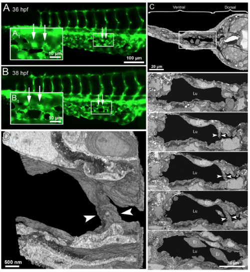
Serial block face electron microscopy (SBF-EM) demonstrating the 3D ultra-structure of newly formed intussusceptive pillar. (A) In vivo GFP images of CVP at 36 hpf, revealing intact vascular surface; the site of future pillar formation is indicated by white arrows. (A 1 ) Symbolizes the rectangle in A at higher magnification. (B,B 1 ) At 38 hpf in the area of interest dark dots (indicated by arrows) represent the newly-formed pillars. (C) Overview of the SBF-SEM transverse section of zebrafish embryo (each slice measuring 75 nm thick) shows the dorsal and ventral regions at 38 hpf and the ventral region with the CVP is indicated in the white rectangle. (C 1 –C 4 ) Corresponding to CVP serial sections (650, 677, 682, 690 and 693) illustrating the ultra-structure of a trans-luminal intussusceptive pillar (indicated by arrowheads). The other structures in the CVP like endothelial cells (EC), erythrocytes (Er) and vascular lumen (Lu) are respectively indicated. (D) Represents the 3D reconstruction of intussusceptive pillar (indicated with large arrowheads) corresponding to the proximal one in B1 and to the C1–C4 SBF-SEM sections. For further information, see supplementary Movie S4. |