
FIGURE
Fig. S2
- ID
- ZDB-FIG-180821-12
- Publication
- Keßler et al., 2018 - A zebrafish model for FHL1-opathy reveals loss-of-function effects of human FHL1 mutations
- Other Figures
- All Figure Page
- Back to All Figure Page
Fig. S2
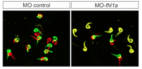
Spontaneous movement assay with false-colored superimposed overviews of 24 hpf control of control-injected embryos and of the fhl1a-morphants. 85% ± 3% (n = 20 embryos per experiment, three independent experiments) of wild-type embryos moved spontaneously within a timeframe of 10 seconds. In contrast, only 22% ± 2% (n = 20 embryos per experiment, three independent experiments, p < 0.0001) of the fhl1a-morphants exhibited spontaneous movement. |
Expression Data
Expression Detail
Antibody Labeling
Phenotype Data
| Fish: | |
|---|---|
| Knockdown Reagent: | |
| Observed In: | |
| Stage Range: | Prim-5 to Protruding-mouth |
Phenotype Detail
Acknowledgments
This image is the copyrighted work of the attributed author or publisher, and
ZFIN has permission only to display this image to its users.
Additional permissions should be obtained from the applicable author or publisher of the image.
Full text @ Neuromuscul. Disord.