
Fig. 4-S1
- ID
- ZDB-FIG-180808-32
- Publication
- Kendall et al., 2018 - PAX3-FOXO1 transgenic zebrafish models identify HES3 as a mediator of rhabdomyosarcoma tumorigenesis.
- Other Figures
- All Figure Page
- Back to All Figure Page
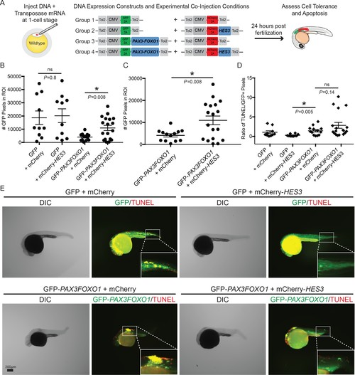
HES3 facilitates cellular tolerance of CMV-PAX3FOXO1 expression in developing zebrafish but does not alleviate the apoptosis phenotype. (A) Schematic of the strategy to assess the impact of human HES3 expression on PAX3-FOXO1 cell tolerance and apoptosis in wildtype developing zebrafish. The CMV promoter was utilized in a mosaic injection strategy, and the experimental groups included the following combinations of two independent plasmids: (1) GFP + mCherry, (2) GFP + mCherry-HES3, (3) GFP-PAX3FOXO1 + mCherry, and (4) GFP-PAX3FOXO1 + mCherry-HES3. Concentrations of injected plasmids were an equivalent molarity to 25 ng/µL of CMV-GFP2A-PAX3FOXO1 (B) Quantification of the number of GFP positive pixels for each embryo imaged at 24 hr post-fertilization using the same settings. Each marker represents a single zebrafish embryo, n = 10–18 embryos per group. Black bar is the mean, error bars represent SEM, and * indicates p<0.05, two-tailed Student’s t-test, ns- not significant. ROI- region of interest. (C) Same samples as in B plotted for the PAX3-FOXO1 and PAX3-FOXO1 + HES3 injection groups. (D) Quantification of TUNEL-positive pixels normalized to GFP-positive pixels indicates that HES3 is not inhibiting PAX3-FOXO1-induced apoptosis at 24 hr post fertilization. Black bar is the mean, error bars represent SEM, n = 10–18 embryos per group, * indicates p<0.05, two-tailed Student’s t-test. ns- not significant. (E) Representative images from the four injection groups: (1) GFP + mCherry, (2) GFP + mCherry-HES3, (3) GFP-PAX3FOXO1 + mCherry, and 4) GFP-PAX3FOXO1 + mCherry-HES3. Embryos were fixed at 24 hr post-injection, TUNEL (rhodamine) performed, and then embryos were counter-stained for GFP to denote transgene expression. |