|
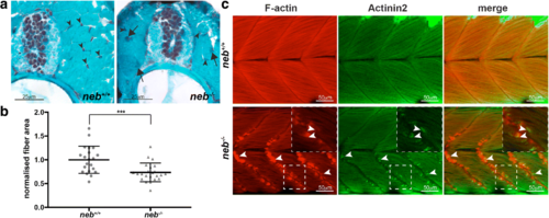
Characterisation of skeletal muscle pathology in neb−/− fish. a Gomori trichome staining of neb−/− skeletal muscle sections reveal the presence of dark regions (arrows) throughout the muscle indicative of nemaline bodies not observed in neb +/+ fish. Nuclei (arrowhead) are evenly organized in neb +/+ , however, appear disorganized in neb−/− fish. b Quantification of normalized fiber area from Gomori trichome stained sections in neb−/− (n = 23 fibers) compared to neb+/+ fish (n = 21 fibers). Error bars represent mean±SD, *** p < 0.001. c neb−/− mutants exhibit F-actin (red) and Actinin2 (green) positive aggregates at the myosepta (arrowheads) (and zoomed inset) compared to wildtype siblings at 2 dpf
|