
Fig. 7
- ID
- ZDB-FIG-180727-33
- Publication
- Eno et al., 2018 - Slow calcium waves mediate furrow microtubule reorganization and germ plasm compaction in the early zebrafish embryo
- Other Figures
- All Figure Page
- Back to All Figure Page
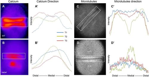
FMA distal enrichment correlates with SCW directionality. (A-B′) Calcium levels in single Tg[βactin2:GCaMP6s] transgenic embryos during furrow progression, with plotted profiles (A′,B′) corresponding to boxed areas. Wild-type embryos (A′), but not nebel mutants (B′), show increasing distal calcium levels. (C-D′) Microtubules in EMTB::EGFP transgenic embryos during furrow progression (C,D), with plotted profiles (C′,D′). Wild-type embryos (C′), but not nebel mutants (D′), show distal enrichment. Time points T1, T2 and T3 correspond, respectively, to frames within one-tenth of the total sequence of acquired images spanning a furrow formation cycle at the beginning, middle and end of that cycle. See Fig. S4 for additional profiles. |
| Fish: | |
|---|---|
| Observed In: | |
| Stage Range: | 2-cell to 4-cell |