
FIGURE
Fig. 4
- ID
- ZDB-FIG-180717-4
- Publication
- Yoo et al., 2018 - Impact of Nicotine Exposure on Hair Cell Toxicity and Embryotoxicity During Zebrafish Development
- Other Figures
- All Figure Page
- Back to All Figure Page
Fig. 4
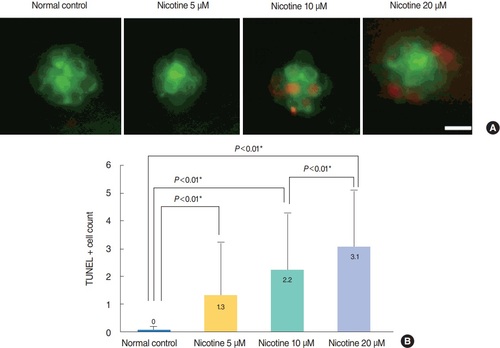
Nicotine-induced apoptosis in hair cells within neuromasts. Nicotine-induced apoptosis was confirmed by terminal deoxynucleotidyl transferase-mediated dUTP-biotin nick end labeling (TUNEL) assay. (A) Green dots represent hair cells. Apoptotic cells appear as light red dots in the merged image obtained by fluorescent microscopy after the TUNEL reaction. All images were obtained using transgenic (brn3c:EGFP) zebrafish at 120 hours post-fertilization. Scale bar=10 μm (×40). (B) The average number of TUNEL-positive cells was measured in each neuromast (supraorbital [SO1, SO2], otic [O1], and occipital [OC1]; total n=50). At a concentration of 40 μM, the fish were almost all dead and statistical analysis was unavailable. The number of TUNEL-positive cells was significantly increased in nicotine-exposed groups (P<0.001, one-way analysis of variance). Only statistically significant pair-wise comparisons in post-hoc analysis are shown. *Statistically significant (P<0.05).
|
Expression Data
Expression Detail
Antibody Labeling
Phenotype Data
Phenotype Detail
Acknowledgments
This image is the copyrighted work of the attributed author or publisher, and
ZFIN has permission only to display this image to its users.
Additional permissions should be obtained from the applicable author or publisher of the image.
Full text @ Clin. Exp. Otorhinolaryngol.