
Fig. 4
- ID
- ZDB-FIG-180717-25
- Publication
- Schott et al., 2018 - EmbryoMiner: A new framework for interactive knowledge discovery in large-scale cell tracking data of developing embryos
- Other Figures
- All Figure Page
- Back to All Figure Page
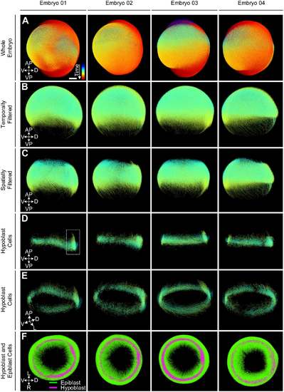
Steps that were performed for extracting hypoblast cells in four different wild-type zebrafish embryos with developmental time ranging from 2–14 hpf (A). The knowledge discovery process was designed interactively on Embryo 01. First, the embryo was filtered temporally (5–7.25 hpf) and spatially (region around the blastoderm margin) to focus on the region of interest (B, C). The two groups of cells were separated using a feature-based clustering approach (D-F). The whole analysis pipeline was then applied to Embryos 02-04 resulting in the extraction of the same internalizing cells in all embryos. The color code in panels (A-E) indicates time from 2–14 hpf and the group association to hypoblast (magenta) or epiblast (green) in panel (F). |