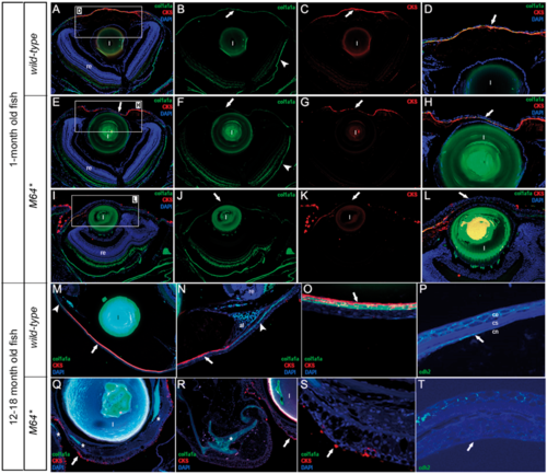
Immunohistochemical analysis of 1-mpf and adult pitx2M64* mutant phenotype. 1-mpf wild-type (A–D) and pitx2M64* mutant (E–L) sections stained with col1a1a (green), CKS (red) and DAPI (blue). D, H and L represent magnified images of the corresponding boxed regions in A, E and I. Wild-type corneal stroma staining reveals a continuous, uniform stromal layer with col1a1a (A, B, D) appearing both in the cornea (arrows in B and D) and around the eye (arrowhead in B) and CKS (A, C, D) being specific to the corneal stroma (arrows in C, D). In mutants, interruptions in or absence of col1a1a (E, F, H–J, L) and CKS (E, G–I, K, L) staining indicates disorganization of the corneal stroma (arrows in E–H, J–L). CKS also detects mucin-secreting goblet cells in the eye periphery and aberrantly approaching the corneal epithelium (I, K, L). Adult wild-type (M–O) and pitx2M64* mutant (Q–S) sections stained with col1a1a (green), CKS (red) and DAPI (blue). Wild-type adult corneal stroma continues to co-express col1a1a and CKS in a continuous layer (arrows in M and O) with CKS localized specifically to the cornea (arrows in M, N) and col1a1a also present around the eye (arrowheads in M, N). In mutant adults, col1a1a and CKS staining is absent from the corneal stroma region (Q). Instead, col1a1a stains ectopic cartilage structures in the endothelial region and ocular periphery (asterisks in Q, R,), whereas CKS stains mucin-secreting goblet cells in both the eye periphery and corneal epithelium (arrows in Q–S). Adult wild-type (P) and pitx2M64* mutant (T) sections stained with N-cadherin (green). In wild-type corneas, cdh2 detects both the corneal epithelium and the monolayer corneal endothelium (arrow in P). In mutants, while cdh2 staining was detectable but less robust in the epithelium, it did not appear in the endothelial region indicating the absence of a defined corneal endothelial layer (arrow in T). al, annular ligament; ce, corneal epithelium; cn, corneal endothelium; cs, corneal stroma; l, lens, re, retina.
|
