
FIGURE
Fig. S8
- ID
- ZDB-FIG-180712-37
- Publication
- Sánchez-Iranzo et al., 2018 - Transient fibrosis resolves via fibroblast inactivation in the regenerating zebrafish heart
- Other Figures
- All Figure Page
- Back to All Figure Page
Fig. S8
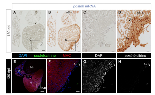
Expression pattern of wt1a:GFP and postnb:citrine in fully regenerated hearts. (A–D) postnb mRNA in situ hybridization (purple) followed by anti-GFP immunohistochemistry (brown) of sections of wt1a:GFP ventricles at 130 dpi (n = 3/3). Arrowheads mark wt1a:GFP+ cells. Sections were stained simultaneously with those shown in Fig. 1 C–F. (E–H) Immunofluorescence with anti-GFP (green) and anti-MHC (red) of a postnb:citrine heart section at 130 dpi (n = 4/4). Nuclei are counterstained with DAPI. Arrowheads mark GFP+ cells. [Scale bars, 100 μm (A, B, and E) and 25 μm (C, D, and F).] |
Expression Data
Expression Detail
Antibody Labeling
Phenotype Data
Phenotype Detail
Acknowledgments
This image is the copyrighted work of the attributed author or publisher, and
ZFIN has permission only to display this image to its users.
Additional permissions should be obtained from the applicable author or publisher of the image.
Full text @ Proc. Natl. Acad. Sci. USA