
Fig. S6
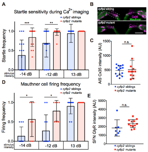
Startle sensitivity and Mauthner firing is increased in cyfip2 mutants during in vivo Ca2+ imaging, Related to Figures 4 and 5 (A) Startle frequency in cyfip2 siblings and mutants during head-restrained Ca2+ imaging following low (-14 dB), medium (-12 dB), and high (13 dB) intensity acoustic stimuli (**p<0.01, ***p<0.001, Mann-Whitney; mean ± SD). (B) Representative examples of Cx35 labeling of the Mcell axon initial segment (AIS). Dashed box indicates area analyzed in (C) (scale bar: 10 μm). (C) Quantification of Cx35 staining on Mcell AIS (n=14 siblings, 12 mutants; p=0.67; mean ± SD). (D) Mcell firing frequency in response to acoustic stimuli was measured by Ca2+ imaging in head-restrained 5 dpf larvae using Tg(Gal4FF- 62A);Tg(UAS:GCaMP6s) (n=11 siblings, 7 mutants; *p<0.05, Mann-Whitney; mean ± SD). (E) Total intensity of GlyR staining on Spiral Fiber Neurons (SFN) was unchanged in cyfip2 mutants (n=7 sibling larvae, 12 mutant larvae; p=0.13, Mann-Whitney; mean ± SD). |