
Fig. 1
- ID
- ZDB-FIG-180627-34
- Publication
- Sedykh et al., 2017 - Zebrafish Rfx4 controls dorsal and ventral midline formation in the neural tube
- Other Figures
- All Figure Page
- Back to All Figure Page
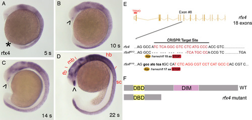
Rfx4 expression and mutagenesis. Representative zebrafish embryos stained using WISH to visualize distribution of rfx4 RNA show expression in the caudal neural tube, including the midbrain, hindbrain, and spinal cord at 5 s (A), 10 s (B), 14 s (C), and 22 s (D). Expression in the preoptic diencephalon is detectable by 10 s and strong by 22 s. There is no detectable expression in the Kupffer's vesicle (KV; asterisk in A). fb, forebrain; mb, midbrain; hb, hindbrain; sc, spinal cord. Arrowheads mark expression in preoptic diencephalon. E: CRISPR assays were used to target a site in the 8th exon of 18 rfx4 exons. Two mutant alleles of rfx4 were generated: uw8013, which encodes a 13‐nt deletion, and uw8110, which encodes a 10‐insertion, at the CRISPR target site. Location of the translation‐blocking morpholino (TBMO) is indicated by a line over the translation start site in exon 1. F: Schematic representation of the wildtype (full‐length) and mutant (truncated) rfx4 proteins. The conserved DNA binding winged‐helix domain (DBD; amino acids 61–136) is indicated in yellow; the conserved dimerization domain (DIM; amino acids 315–487) is shown in purple. The predicted proteins encoded by rfx4uw8013 and rfx4uw8110 are truncated due to frameshifts after amino acid 262; both truncated proteins lack the dimerization domain. |
| Gene: | |
|---|---|
| Fish: | |
| Anatomical Terms: | |
| Stage Range: | 5-9 somites to 20-25 somites |