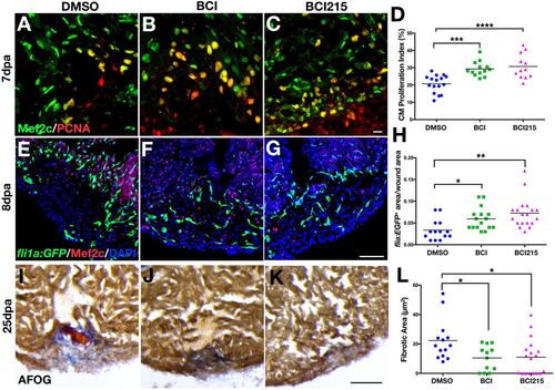
Chemical inhibition of Dusp6 increases cardiomyocyte proliferation and angiogenesis and reduces fibrosis after cardiac injury. (A-C) Adult zebrafish hearts at 7 dpa, injected for 6 days with DMSO (n=16) (A), 0.5 mg/kg BCI (n=13) (B) or 0.5 mg/kg BCI215 (n=12) (C) and stained for Mef2c and Pcna. (D) Quantification of proliferating cardiomyocytes at 7 dpa. BCI and BCI215 increased cardiomyocyte proliferation compared with DMSO vehicle. (E-G) Tg(fli1a:EGFP)y1 hearts at 8 dpa injected for 6 days with BCI (n=16) (F), BCI215 (n=19) (G) or DMSO (n=14) (E) and stained for Mef2c. (H) Quantification of new vessels formed inside the clot area of hearts at 8 dpa. (I-K) Sections of hearts at 25 dpa stained with AFOG to visualize the scar. Intact cardiac muscle stains orange-brown, fibrin stains red and collagen blue. Fish injected with BCI (n=12) (J) and BCI215 (n=18) (K) resolved the injury faster than DMSO-injected fish (n=14) (I). (L) Quantification of clot areas at 25 dpa. (D,H,L) ****P<0.0001, ***P<0.001, **P<0.01, *P<0.05, one-way ANOVA. Scale bars: 100 µm.
|
