
Fig. 4
- ID
- ZDB-FIG-180622-12
- Publication
- Kague et al., 2018 - Zebrafish sp7 mutants show tooth cycling independent of attachment, eruption and poor differentiation of teeth
- Other Figures
- All Figure Page
- Back to All Figure Page
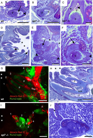
Tooth abnormalities in thesp7mutant. Toluidine blue stained 2 µm sections of teeth in 6 months old wt (A, D and H) and sp7 mutant (B-C, E-F and J) zebrafish. Confocal imaging of teeth in 5 months wt and sp7 -/- zebrafish (G and I, respectively). A) Replacement tooth of a wt, with enameloid (arrowhead) and dentin (d) containing tubules (arrow), with predecessor tooth (asterisk) partly visible, localized in proximity of the pharyngeal epithelium. B) Overview of a defective tooth in the sp7 mutant with a thin layer of dentin (d) and highly cellular pulp (p). C) Magnification of a sp7 mutant tooth. Note small amount of dentin (d) and enameloid with traces of organic matter (arrowhead) but well organized enamel organ with polarized ameloblasts (asterisk). Note densely cellular pulp (p). D-E) Lower magnification picture to show the pharyngeal cavity (asterisks) and tooth arrangement in wt (D) and sp7 mutant (E). In wt, teeth are oriented with tooth tips (arrows) pointing to the pharyngeal cavity (asterisks), while in the sp7 mutant some teeth have an abnormal orientation with their tip (arrow) turned away from the pharyngeal cavity. Pairs of teeth can also be observed (arrowheads). F) A pair of tooth germs (predecessor, arrow and successor, arrowhead). These germs have an orientation relative to each other much as in the wt (compare with A). Note that the densely cellular pulps of both germs are interconnected without sharp boundary. G-J) Confocal imaging (G, I) and histological sections (H, J) of similar teeth to show pulps in wt (G, H) and sp7 mutant (I, J). G, I). wt (G) and sp7 mutant (I) carrying (Tg(RUNX2:egfp) and stained for Alizarin Red S, imaged using confocal microscopy followed by 3D projection. The pharyngeal cavity region (asterisks), pharyngeal bone (b) and bone of attachment (boa, in wt only) are labelled. Numbers label the different teeth. Note that the bone of attachment delimits each pulp (p) in the wt (G) while in sp7 mutants (I) the lack of bones of attachment leads to abnormally connected pulps (p). H, J) Toluidine blue stained 2 µm cross section of pulps (p) in wt (H) and multi-pulps (p) connected in the sp7 mutant (J). The pharyngeal cavity region is indicated by asterisks and the bone of attachment (boa) is labelled in the wt. Scale bars A, B, F and J = 50 µm; C = 20 µm; D, E, G, H and I= 100 µm. |
| Fish: | |
|---|---|
| Observed In: | |
| Stage: | Adult |
Reprinted from Developmental Biology, 435(2), Kague, E., Witten, P.E., Soenens, M., Campos, C.L., Lubiana, T., Fisher, S., Hammond, C., Brown, K.R., Passos-Bueno, M.R., Huysseune, A., Zebrafish sp7 mutants show tooth cycling independent of attachment, eruption and poor differentiation of teeth, 176-184, Copyright (2018) with permission from Elsevier. Full text @ Dev. Biol.