
FIGURE
Fig. S7
Fig. S7
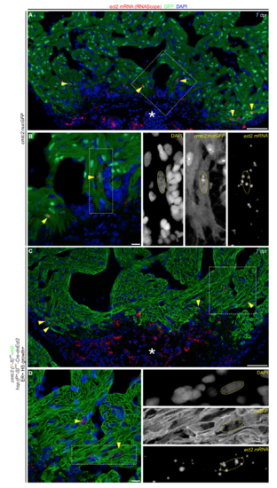
Ect2 is re-expressed after injury in polyploid-enriched hearts, related to Figure 5 and Figure 6. (A,C) RNAScope in situ hybridization showing ect2 transcripts (yellow arrowheads) in a section from the indicated cohorts at 7 dpr, immunostained for GFP and counterstained with DAPI. Asterisk indicates the wound area. (B,D) Magnified regions from (A) and (D). Boxed region is shown to the right with individual fluorescent signals for DAPI, GFP and ect2 mRNA. n=4 hearts per group with 3 sections per heart. Scale bars: 50 μm (A,C), 10 μm (B,D). |
Expression Data
Expression Detail
Antibody Labeling
Phenotype Data
Phenotype Detail
Acknowledgments
This image is the copyrighted work of the attributed author or publisher, and
ZFIN has permission only to display this image to its users.
Additional permissions should be obtained from the applicable author or publisher of the image.
Reprinted from Developmental Cell, 44, González-Rosa, J.M., Sharpe, M., Field, D., Soonpaa, M.H., Field, L.J., Burns, C.E., Burns, C.G., Myocardial Polyploidization Creates a Barrier to Heart Regeneration in Zebrafish, 433-446.e7, Copyright (2018) with permission from Elsevier. Full text @ Dev. Cell