
Fig. 3
- ID
- ZDB-FIG-180501-23
- Publication
- Paffett-Lugassy et al., 2017 - Unique developmental trajectories and genetic regulation of ventricular and outflow tract progenitors in the zebrafish second heart field
- Other Figures
- All Figure Page
- Back to All Figure Page
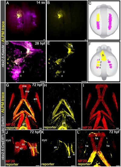
Pharyngeal arch 2 muscles and hypobranchial artery endothelium derive from nkx2.5+ progenitors in the ALPM. (A-F) Confocal z-stacks (A,B,D,E) and schematic diagrams (C,F) of a 14-somite stage (ss) (16 hpf) Tg(nkx2.5:Kaede) embryo immediately following left-side photoconversion of the Kaede+ ALPM (anterior half; A-C) and the same embryo at 28 hpf (D-F) imaged in the green (pseudocolored magenta) and red (pseudocolored yellow) channels. Dorsal views are shown. Anterior is upwards. Merged (A,D) and single-channel (B,E) images are shown. In all five embryos, photoconverted cells traced to the heart and the pharyngeal arches as shown. (G-L) Confocal z-stacks of pharyngeal regions in 72 hpf Tg(nkx2.5:CreERT2), Tg(ubi:Switch) embryos pulsed with 4-OHT between tailbud and 16 ss (10-17 hpf). Animals were double immunostained to detect the mCherry reporter protein and striated muscle (MF20 antibody) before imaging in the red (pseudocolored yellow) and far-red (pseudocolored red) channels, respectively. Ventral (G-I,L) and left lateral (J,K) views are shown. Anterior is upwards (G-I,L) or leftwards (J,K). All 12 animals exhibited lineage tracing to the described structures. Numbers label the pharyngeal arches. h, heart. Ventral pharyngeal arch (PA) 1 (mandibular) muscle: ima, intermandibular anterior. Middle PA1 muscles: imp, intermandibular posterior; am, adductor mandibulae. Dorsal PA1 muscles: lap, levator arcus palatine; do, dilator operculi. Ventral PA2 (hyoid) muscle: ih, interhyal. Middle PA2 muscle: hh, hyohyal. Dorsal PA2 muscles: ah, adductor hyomandibulae; ao, adductor operculi; lo, levator operculi. Vessel: ha, hypobranchial artery. Cardiac structures: oft, outflow tract; v, ventricle. Scale bars: 25 µm. |