
FIGURE
Fig. 1
- ID
- ZDB-FIG-180424-14
- Publication
- Silvent et al., 2017 - Zebrafish skeleton development: High resolution micro-CT and FIB-SEM block surface serial imaging for phenotype identification
- Other Figures
- All Figure Page
- Back to All Figure Page
Fig. 1
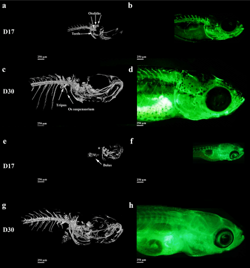
Micro-CT-scan and 3D-reconstructions (a,c,e,g) and fluorescence microscopy after calcein staining imaging (b,d,f,h) of side views of calcified skeletal structures in wild type (a,b,c,d) and nacre (e,f,g,h) zebrafish larvae at 17 dpf (a, b, e, f) and 30 dpf (c,d,g,h). No vertebra is observed using the CT scan observation at D17 (asterisk). |
Expression Data
Expression Detail
Antibody Labeling
Phenotype Data
| Fish: | |
|---|---|
| Observed In: | |
| Stage: | Days 14-20 |
Phenotype Detail
Acknowledgments
This image is the copyrighted work of the attributed author or publisher, and
ZFIN has permission only to display this image to its users.
Additional permissions should be obtained from the applicable author or publisher of the image.
Full text @ PLoS One