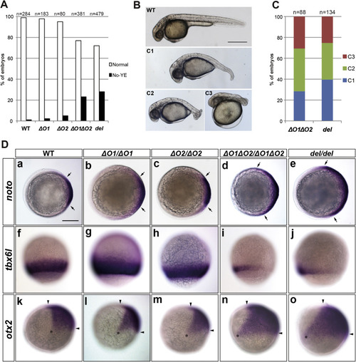
Role of ORF1 and ORF2 in the zygotic wnt8a-mediated development of caudal and ventrolateral tissues. (A) Percentage of mutants showing a lack of yolk extension (no yolk extension: No-YE). Embryos were obtained by crossing WT, or wnt8aΔO1(ΔO1), wnt8aΔO2 (ΔO2), wnt8aΔO1ΔO2 (ΔO1ΔO2), or wnt8adel (del) heterozygote pairs. Phenotypes were observed at 30 hpf. The number of observed embryos is indicated. (B) WT and wnt8a mutant phenotypes. Lateral views with anterior to the left. The No-YE embryos were categorized into three classes. C1 embryos had a slightly shortened and ventrally bent tail, C2 embryos had a short and curled tail, and C3 embryos had an enlarged head and a truncated tail. This phenotype classification is essentially the same as one described in a previous publication (Shimizu et al., 2005), except for being based on the No-YE phenotype. (C) Proportion of No-YE embryos showing each phenotype class. The number of observed embryos is indicated. (D) Expression of noto (formerly called floating head, shield stage, a-e), tbx6l (tbx6, 80% epiboly stage, f-j), and otx2 (100% epiboly stage, k-o) in WT (a, f, k) and ΔO1 (b, g, l), ΔO2 (c, h, m), ΔO1ΔO2 (d, i, n), and del (e, j, o) homozygous mutants. Animal pole views with dorsal to the right (a-e). Lateral views with dorsal to the right (f-o). noto, tbx6l, and otx2 are markers of the axial mesoderm, the paraxial mesoderm, and the rostral neuroectoderm (forebrain and midbrain), respectively. The lateral limits of noto expression are marked by arrows, the rostral and caudal limits of otx2 expression are marked by arrowheads, and the ventral limit of otx2 expression is marked by an asterisk. The noto expression was not greatly affected in the ΔO1 and ΔO2 single mutants, but it was prominently expanded in the ΔO1ΔO2 and del mutants (ΔO1, n=0/86; ΔO2, n=0/94; ΔO1ΔO2, n=10/42; del, n=15/44 in embryos from the heterozygous crosses; ΔO1, n=0/7; ΔO2, n=0/7; ΔO1ΔO2, n=4/4; del, n=6/6 in genotyped homozygotes). The tbx6l expression was not affected in the ΔO1 or ΔO2 mutants, but was reduced or absent in the ΔO1ΔO2 and del mutants (ΔO1, n=0/353; ΔO2, n=0/96; ΔO1ΔO2, n=27/83; del, n=37/140 in embryos from the heterozygous crosses; ΔO1, n=0/22; ΔO2, n=0/9; ΔO1ΔO2, n=11/11; del, n=10/10 in the genotyped homozygotes). The otx2 expression was not affected in the ΔO1 or ΔO2 mutants, but was expanded in the ΔO1ΔO2 and del mutants (ΔO1, n=0/73; ΔO2, n=0/99; ΔO1ΔO2, n=10/49; del, n=18/57 in embryos from the heterozygous crosses; ΔO1, n=0/6; ΔO2, n=0/7; ΔO1ΔO2, n=6/6; del, n=6/6 in genotyped homozygotes). Scale bars: 500 µm in B; 200 µm in D.
|
