
Fig. 8
- ID
- ZDB-FIG-180411-12
- Publication
- Veil et al., 2017 - Maternal Nanog is critical for the zebrafish embryo architecture and for cell viability during gastrulation
- Other Figures
- All Figure Page
- Back to All Figure Page
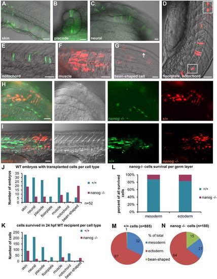
Transplantation experiment reveals cell-autonomous death of nanog−/− cells. (A-G) Scored cell types: skin cells (A), placode, e.g. eye (B), neural epithelial cells in the hindbrain (C), floorplate cells (D), cells in the notochord (E), muscle cells (F), non-specified ‘bean-shaped’ cells (G). Anterior is always to the right, except in D in which anterior is toward the top. (H,I) Two examples of differentiated co-transplanted wild-type (+/+) and nanog−/− cells in 24 hpf wild-type (WT) host. Overlay, differential interference contrast, green and red channels from left to right. (H) Neural tissue: nanog−/− cells (green) differentiated as skin and +/+ cells (red) contributed to neural epithelium in the hindbrain region of the 24 hpf WT host. (I) Trunk muscles: 6 nanog−/− (red) muscle fibers and 27 +/+ (green) muscle fibers are visible. (J) Number of embryos with at least one transplanted cell per cell type for +/+ and nanog−/− cells in 52 hosts (note that the embryos which contained several types of donor cells were scored more than once). (K) Total number of transplanted cells per tissue in 52 hosts. P-values for the difference between the numbers of +/+ and nanog−/− cells were significant for all tissues (P<0.01, paired t-test; see also Table S3). (L) Percentage of surviving cells separated by germ layer: mesoderm (muscle and notochord) and ectoderm (skin, neural cells, placodes and floorplate cells). Contribution of nanog−/− cells to both mesodermal and ectoderm derivatives was <20%. (M,N) Distribution of +/+ and nanog− /− cells between mesodermal and ectodermal tissue and non-specified ‘bean-shaped’ cells. Scale bars: 40 µm (A,C-G,I); 200 µm (B); 100 µm (H). |