
FIGURE
Fig. s3
- ID
- ZDB-FIG-180410-18
- Publication
- Wu et al., 2017 - Patient-derived xenograft in zebrafish embryos: a new platform for translational research in gastric cancer
- Other Figures
- All Figure Page
- Back to All Figure Page
Fig. s3
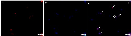
Fluorescent microscopy analysis of dissociated embryos. Xenografted embryos were dissociated and the resulting cell suspension were analyzed by fluorescent microscopy. The eight cells in the field of view that stain positive for CM-DiI colocalize with individual nuclei (white arrows) stained with DRAQ5 nuclear stain. |
Expression Data
Expression Detail
Antibody Labeling
Phenotype Data
Phenotype Detail
Acknowledgments
This image is the copyrighted work of the attributed author or publisher, and
ZFIN has permission only to display this image to its users.
Additional permissions should be obtained from the applicable author or publisher of the image.
Full text @ J. Exp. Clin. Cancer Res.