|
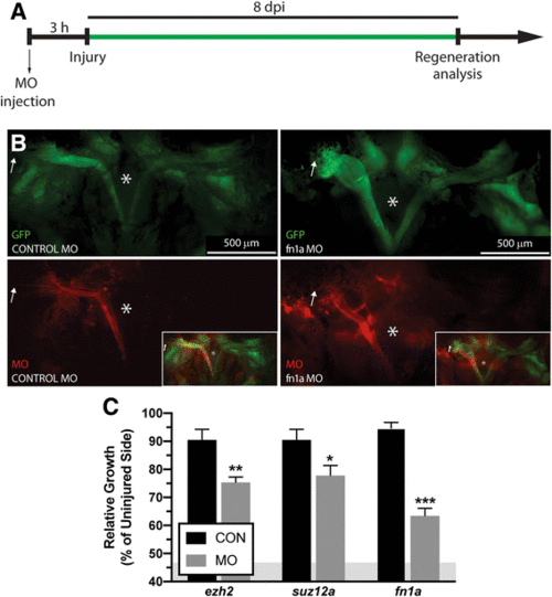
Regeneration following knockdown of select differentially expressed genes. a LR regeneration assays were performed according to the schematic. b Craniectomy was performed to visualize EGFP-labeled muscle at 8 dpi. Brain was removed to allow visualization of the skull base (*) Microinjected MOs are detected throughout the entire regenerating muscle, including the distal ends (white arrow). Control MO (left) and fn1a MO (right) injected fish are pictured, Additional file 26: Figure S14 shows representative examples of injected fish with MO targeting the rest of the genes. c Quantification of LR regeneration at 8 dpi, all MOs targeting specific mRNA decreased muscle regeneration at 8 dpi compared to control MO injected fish. Values are averages ± SEM (n = 5–7) in control MO or target gene MO injected fish. *P < 0.05; **P < 0.01; *** P < 0.001; Student’s t-test. The residual muscle left following myectomy surgery (46.77 ± 4.8%, average ± SD) is represented as a grey area in C
|