
Fig. 5
- ID
- ZDB-FIG-180403-38
- Publication
- Furlan et al., 2017 - Life-Long Neurogenic Activity of Individual Neural Stem Cells and Continuous Growth Establish an Outside-In Architecture in the Teleost Pallium
- Other Figures
- All Figure Page
- Back to All Figure Page
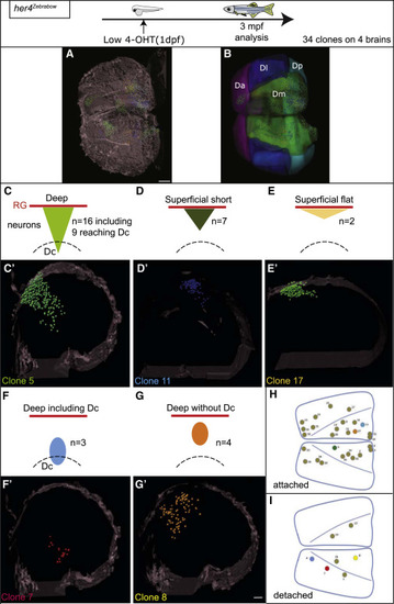
Individual Pallial RGs Are Neurogenic Lifelong Top: experimental design. (A) Whole-mount dorsal view (anterior left) of a 3-mpf pallium from a her4Zebrabow,4OHT(1dpf) fish, cleared and scanned. The position of individual cells of each clone is plotted (color coded). Brain surface (gray) was digitally added to help visualization. Scale bar, 150 μm. (B) Corresponding location of pallial subdivisions. (C–G′) Schematics (C–G) (red lines: RG layer; dashed lines: Dc limit) and thick optical cross-sections (C′–G′) of brains as in (A) highlighting the different categories of attached clones (C–E′) or detached clones (F–G′) (Table S1). “Deep” versus “superficial” clones contain neurons born before or at 5 dpf (Table S2Table S2). (F–G′) Schematics (F and G) and thick optical cross-sections (F′ and G′) of brains as in (A) with examples of detached clones. (H and I) Position of the 34 clones attached (H) or detached (I) on flattened dorsal views of the 3-mpf pallium. Clones illustrated in (C′)–(G′) are colored. Scale bar, 80 μm. See also |