
Fig. 3
- ID
- ZDB-FIG-180403-12
- Publication
- Posner et al., 2017 - The zebrafish as a model system for analyzing mammalian and native α-crystallin promoter function.
- Other Figures
- All Figure Page
- Back to All Figure Page
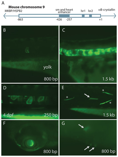
Mouse αB-crystallin promoter fragments produced native expression in zebrafish embryos. Enhancer elements of a promoter upstream of mouse αB-crystallin were previously shown to regulate expression in skeletal muscle (sm), heart and lens (lsr1 and 2) (A; adapted from Swamynathan & Piatigorsky, 2002). Fragments containing 250 bp, 0.8 and 1.5 kb lengths of this promoter produced GFP expression in zebrafish embryo notochord (B–D), skeletal muscle (E), lens (F) and heart (G; arrows). (E) shows GFP expression in both fast (noted by *) and slow twitch (noted by arrows) muscle fibers. The yolk remaining in these embryos is autofluorescent. The 250 bp fragment, which lacked the heart and skeletal muscle enhancer, produced less frequent GFP expression in these tissues, and GFP expression onset was slightly earlier (Tables 3 and 4). |