
Fig. 7
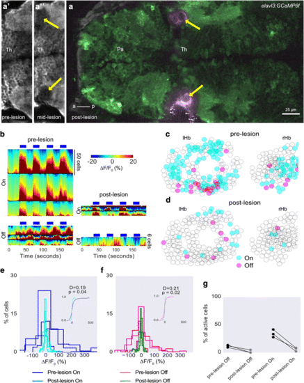
The effect of lesioning the anterior thalamic neuropil on habenula response to light. a Dorsal view of an elavl3:GCaMP6f fish, showing lesion bubbles in the anterior thalamic neuropil created by a femtosecond laser (arrows). The bubble reflects the two-photon laser, and is thus captured in a separate channel from GCaMP6f fluorescence. a’, a’’ Close-up of the anterior thalamus neuropil before (a’) and during (a’’) the lesion. The cavity has not yet formed. b Heatmap showing habenula cell responses before (left) and after (right) lesioning in three fish. The scale of the bottom right panel is different from the others as indicated. c, d The cells segmented from all three fish are drawn as circles and overlaid. Responding cells before and after the lesion are colored as indicated. e, f Histogram showing distribution of mean intensity in habenula neurons during light ON (e) and OFF (f) before and after the lesion. Insets show cumulative distribution from all fish. P values and test statistic were obtained using the Kolmogorov–Smirnov test. g Comparison of percentage of cells responding to light ON and OFF before and after the lesion. Each circle is one fish and lines join data points from the same fish. Th Thalamus, Pa Pallium, lHb left habenula, rHb right habenula, a anterior, p posterior. Images are all single optical sections |