
FIGURE
Fig. 4
- ID
- ZDB-FIG-180320-8
- Publication
- Suarez-Bregua et al., 2017 - Targeted Pth4-expressing cell ablation impairs skeletal mineralization in zebrafish
- Other Figures
- All Figure Page
- Back to All Figure Page
Fig. 4
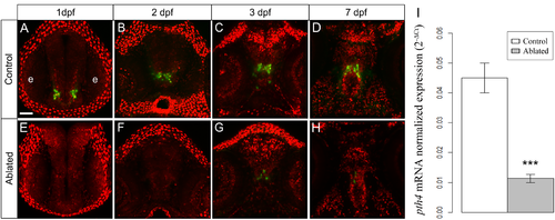
Illustration showing the development of the two clusters of Pth4:eGFP neurons in control (A-D) and ablated larvae (E-H) from 1 to 7 dpf. After first and second round of ablation, 1 and 2 (at 1 and 2 dpf, respectively) the eGFP-expressing cells are totally eliminated (E and F); at 3 dpf and 7 dpf a low number of Pth4:eGFP-expressing neurons recover (G-H), but there remains significantly decreased pth4 gene expression (***p< 0.001) in 7 dpf ablated larvae (I). Results normalized to actb1 are expressed as mean ± SEM. Confocal z-stack projections are shown. Abbreviation: e, eye. Scales bars: 50 μm. |
Expression Data
| Gene: | |
|---|---|
| Fish: | |
| Condition: | |
| Anatomical Term: | |
| Stage: | Days 7-13 |
Expression Detail
Antibody Labeling
Phenotype Data
| Fish: | |
|---|---|
| Condition: | |
| Observed In: | |
| Stage: | Days 7-13 |
Phenotype Detail
Acknowledgments
This image is the copyrighted work of the attributed author or publisher, and
ZFIN has permission only to display this image to its users.
Additional permissions should be obtained from the applicable author or publisher of the image.
Full text @ PLoS One