
Fig. 1
- ID
- ZDB-FIG-180223-9
- Publication
- Teng et al., 2017 - Requirement for Jagged1-Notch2 signaling in patterning the bones of the mouse and human middle ear
- Other Figures
- All Figure Page
- Back to All Figure Page
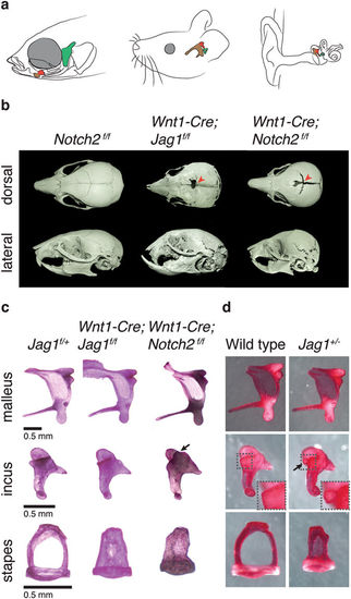
Craniofacial and middle ear defects in mice deficient for Jag1 or Notch2. (a) Diagrams of the heads of zebrafish, mouse, and human (left to right) show homology between the fish jaw skeleton and the mammalian middle ear ossicles. The fish hyomandibula is homologous to the mammalian stapes (green); the fish palatoquadrate is homologous in part to the mammalian incus (red); and the proximal portion of the fish Meckel’s cartilage is homologous to the mammalian malleus (brown). (b) Micro-CT scans of mouse skulls at three weeks of age. Compared to control Notch2 f/f mice, Wnt1-Cre; Jag1 f/f and Wnt1-Cre; Notch2 f/f mice exhibit a persistent foramen (arrowheads in dorsal view) and midfacial hyperplasia resulting in an abnormally shaped skull and malocclusion (lateral view). (c,d) Dissected middle ear ossicles of three-week-old mice stained with Alizarin Red S. Compared to Jag1 f/+ controls, Wnt1-Cre; Jag1 f/f and Wnt1-Cre; Notch2 f/f mice display a fully penetrant columellar stapes phenotype. Wnt1-Cre; Notch2 f/f mice also rarely display an ectopic process from the anterior medial edge of the incus (arrow). Compared to wild-type siblings, some Jag1 +/− mice display a columellar stapes and a small ectopic process from the posterior medial edge of the incus body (arrow and inset). |