
Fig. 1
- ID
- ZDB-FIG-180201-9
- Publication
- Daly et al., 2017 - A Brain-Derived Neurotrophic Factor Mimetic Is Sufficient to Restore Cone Photoreceptor Visual Function in an Inherited Blindness Model
- Other Figures
- All Figure Page
- Back to All Figure Page
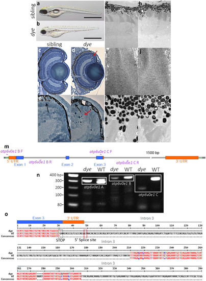
dye mutants display aberrant retinal morphology. Representative lateral and dorsal views in brightfield of sibling larvae (a,a′) and dye mutants (b,b′), displaying pigmentation and gross morphological defects present in the dye mutant, including swim bladder deflation compared to sibling (*in a) and pericardial oedema (black arrow in b). Transverse sections of the retina and ciliary marginal zone of sibling larvae (c,e) and dye mutants (d,f), pyknotic nuclei (red arrows in d,f) present in the ciliary marginal zone indicated dying cells. Transmission electron micrographs of photoreceptors in sibling larvae (g) and dye mutants (h) displaying shortened photoreceptor OS and aberrant morphology despite preserved ultrastructure (i,j). RPE cells in dye mutants are hypopigmented and contain large inclusion bodies compared to sibling larvae (k,l), and the RPE fails to interdigitate with photoreceptors in dye compared to siblings (N = 1 for sibling, N = 3 for dye mutants). (m–o) The mutation in dye affects the zebrafish atp6v0e1 gene. (m) Schematic representation of the exon-intron structure of atp6v0e1, purple lines indicate the annealing position of genotyping primers used in panel n. (n) 1% agarose gel of PCR products from different regions of the atp6v0e1 gene, a 180 bp deletion (white box, n) is detected in the region containing the terminal end of coding exon 3, the full length agarose gel image is included in the supplementary information (Supplementary Fig. S3a). (o) Sequencing of gel extracted PCR products and pairwise sequence alignment to the reference genome sequence (assembly GRCz11) revealed that the deletion removes the stop codon in, and the 3′ UTR encoded in exon 3 and of the GT splice donor site encoded at the exon 3-intron 3 border. |
| Fish: | |
|---|---|
| Observed In: | |
| Stage: | Day 5 |