
Fig. 7
- ID
- ZDB-FIG-180117-37
- Publication
- Ng Chi Kei et al., 2017 - Fate bias during neural regeneration adjusts dynamically without recapitulating developmental fate progression
- Other Figures
- All Figure Page
- Back to All Figure Page
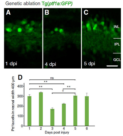
Following genetic ablation, new horizontal and amacrine cells can be observed prior to the proliferative wave. a-c) Micrographs of retinal sections in Tg(ptf1a:GFP) larvae at different days post injury (dpi). d Quantification shows an initial reduction and subsequent increase in the number of Ptf1a:GFP labelled inhibitory neurons. At 3 and 4 dpi, the number is significantly lower (* p-value = 0.018, ** p-value ≤0.01) compared to 1 dpi (baseline) or 5 dpi (regenerated), which are not significantly different from each other (p-value = 0.50). Ns: not significant (p-value >0.05). Results are mean ± SEM. INL: inner nuclear layer; IPL: inner plexiform layer; GCL: ganglion cell layer. Scale bar C = 50 μm |