
Fig. S4
- ID
- ZDB-FIG-180104-9
- Publication
- Djenoune et al., 2017 - The dual developmental origin of spinal cerebrospinal fluid-contacting neurons gives rise to distinct functional subtypes
- Other Figures
- All Figure Page
- Back to All Figure Page
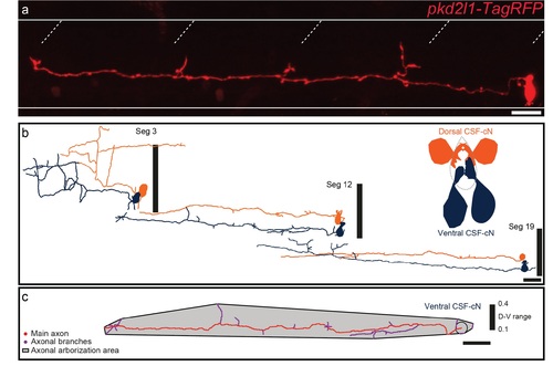
Characterization of CSF-cNs axonal morphology (a) Projection obtained from z-stacks of a single CSF-cN in a wild-type larva previously injected with (pkd2l1-TagRFP). (b) The reconstruction of 3 ventral CSF-cNs (dark blue) and 3 dorsal CSFcNs (orange) from segment (Seg) 3, 12 and 19 shows that, along the rostrocaudal axis, ventral CSF-cNs are morphologically different from dorsal ones. (c) Illustration of the reconstruction of one ventral CSF-cN displays the area covered by the axon and the axonal arborization nomenclature. Horizontal lines represent the limits of the spinal cord and slash dashed lines represent somite boundaries. Vertical black bars represent the dorso-ventral limits of the spinal cord, where the ventral edge corresponds to 0 and the dorsal edge to 1. Scale bars = 20 μm. |