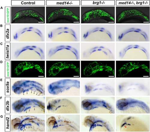
Defects in skeletogenic neural crest differentiation in the jaw forming area of med14 and brg1 mutant embryos. a to d Neural crest cells migrated to the oral ectoderm in both control and mutant embryos. a Snapshot of migrating neural crest cells (marked by sox10:EGFP transgene) in control and mutant embryos at 15 somite stage. b and c The migrating neural crest expressed dlx2a and twist1a at 18 somite stage. d At 24 hpf, neural crest cells reached the brachial arches forming region and condensed. a and d: lateral view with dorsal to top and anterior to left. e to g The mutants showed mis-expression of genes involved in mesenchymal condensations and chondrocyte differentiation. b, c, e, f and g RNA in situ hybridization is shown for expression of dlx2a, tiwst1a, sox9a, dlx3b and hand2. At least 15 embryos for each genotype were analyzed and representative samples are shown. Hollow arrowheads indicate pharyngeal arches; red arrowhead in g indicates the heart tube of embryo. Lateral views with anterior to the left. Scale bars, 100 um
|
