
FIGURE
Fig. 3
- ID
- ZDB-FIG-171027-37
- Publication
- Mathuru, 2016 - Conspecific injury raises an alarm in medaka
- Other Figures
- All Figure Page
- Back to All Figure Page
Fig. 3
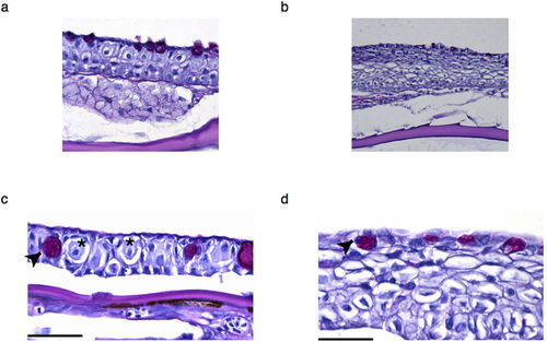
Histology of epidermis of medaka and zebrafish. Low (a,b) and high magnification (c,d) images of the zebrafish (a,c) and medaka (b,d) epidermis tissue. Club cells in the zebrafish epidermis (c) are negative for PAS staining and are indicated by an asterisk (*). Arrowheads show another type of secretory cell that is positive for PAS staining - the mucous goblet cell. Scale bar is 50 um. |
Expression Data
Expression Detail
Antibody Labeling
Phenotype Data
Phenotype Detail
Acknowledgments
This image is the copyrighted work of the attributed author or publisher, and
ZFIN has permission only to display this image to its users.
Additional permissions should be obtained from the applicable author or publisher of the image.
Full text @ Sci. Rep.