
Fig. 4 S1
- ID
- ZDB-FIG-171004-6
- Publication
- Boothe et al., 2017 - A tunable refractive index matching medium for live imaging cells, tissues and model organisms
- Other Figures
- All Figure Page
- Back to All Figure Page
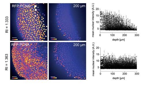
Improved nuclear segmentation in deep tissue layers by Iodixanol supplementation. four hpf zebrafish embryos expressing RFP-PCNA. Top row: Maximum projection (left) and optical XY-section at 200 µm depth (right) of an embryo in regular media (RI = 1.333) highlights a progressive loss of nuclear signal detection and segmentation beyond 100 µm imaging depth, which is quantified in the scatter plot of mean nuclear intensity versus depth (right). Bottom row: RI tuning with Iodixanol to RI = 1.363 enables nuclear detection and segmentation at up to 300 µm imaging depth. Panel order and imaging conditions exactly as above. Scale bars = 50 µm. The colour scheme encodes relative intensity (brightest = white). |