
Fig. 6
- ID
- ZDB-FIG-171002-12
- Publication
- Fernández et al., 2017 - Optimizing pulse compressibility in completely all-fibered Ytterbium chirped pulse amplifiers for in vivo two photon laser scanning microscopy
- Other Figures
- All Figure Page
- Back to All Figure Page
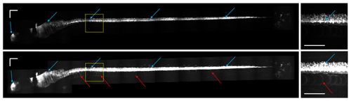
Two-photon mCherry (red) fluorescence from a 5 days old zebrafish larva. The left images are composed of 20 frames of 300x300 µm2 each. The scale bars correspond to 100 µm. The upper left image shows the red fluorescence from mCherry-labelled cells, with the contrast set such that only the brightest cells (central nervous system cells, blue arrows) are slightly saturated. The lower left image has the brightness setting increased, such that finer structures with weaker fluorescence, for example from the cell walls of the notochord (red arrows), just below the bright spinal cord, can be seen. The upper and lower right images show a zoom of the dashed boxes indicated in the respective left images. |