
Fig. 2
- ID
- ZDB-FIG-170922-45
- Publication
- Scahill et al., 2017 - Loss of the chromatin modifier Kdm2aa causes BrafV600E-independent spontaneous melanoma in zebrafish
- Other Figures
- All Figure Page
- Back to All Figure Page
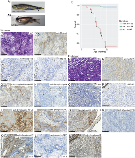
Kdm2aa-deficient fish develop melanoma. Aberrant melanocytic pigmentation on the tail (Ai, arrow) and eye tumours (Aii, arrow) are observed in Kdm2aa-deficient fish. (B) Survival graph for kdm2aasa898 showing incidence of suspected cancer. Each (*) indicates a single culled fish due to suspected cancer. No wild-type or heterozygous siblings developed any suspected cancers. (C) H and E stained section of a tail tumour showing a biphasic appearance with pseudoglandular features (arrow shows an example) alternating with areas of spindle cell growth. (D) Section of tail tumour staining positive for the melanoma marker Melan-A (brown). (E) Section of tail tumour negative for the melanoma marker S100. (F) Section of tail tumour staining negative for the melanoma marker HMB-45. (G) Section of tail tumour staining positive for mitotic marker phospho-histone H3 (brown). (H) Section of tail tumour showing negative staining for the neuroendocrine marker Synaptophysin. Arrow points to example of pseudoglandular structures. (I) Section of tail tumour showing positive staining for the epithelial marker Cytokeratin (brown) in both the pseudoglandular and spindle cell elements. (J) Section of tail tumour with positive phospho-ERK staining (brown). (K) Section of tail tumour with positive phospho-AKT staining (brown) (L) Section of a second tail tumour which did not stain positive for phospho-AKT. (M) H and E stained section from an eye tumour revealing spindle cell morphology. (N) Section of eye tumour staining positive for Melan-A (brown). (O) Eye tumour section showing negative staining for the S100. (P) Section of eye tumour negative for HMB-45. (Q) Section of eye tumour staining positive for phospho-histone H3 (brown). (R) Phospho-ERK staining of eye tumour sections was negative. (S) Section of eye tumour showing positive staining for phospho-AKT (brown). Scale bar in C-S is 100 μm. |
| Fish: | |
|---|---|
| Observed In: | |
| Stage: | Adult |