
Fig. 5
- ID
- ZDB-FIG-170921-38
- Publication
- Kara et al., 2017 - miR-27 regulates chondrogenesis by suppressing Focal Adhesion Kinase during pharyngeal arch development
- Other Figures
- All Figure Page
- Back to All Figure Page
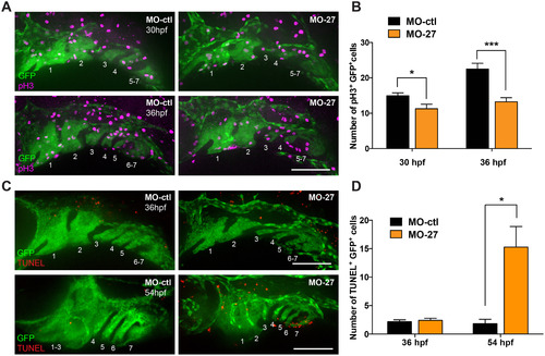
miR-27 knock-down impairs proliferation and survival of PCC cells. (A) Anti-phospho histone 3 (pH3) and anti-GFP immunostaining in Tg(fli1a:eGFP)y1 embryos at 32 and 36 hpf. Embryos were injected with either 5 ng MO-ctl or MO-27 at the single cell stage. Lateral view with anterior to the left. (B) Quantification of pH3+GFP+ cells normalized to the GFP+ area in each embryo. For 30 hpf, n=7; for 36 hpf, n=8. (C) TUNEL staining and anti-GFP immunostaining in Tg(fli1a:eGFP)y1 embryos at 36 and 54 hpf. Embryos were injected with the corresponding morpholinos and image layouts are as described above. (D) Quantification of TUNEL+GFP+ cells normalized to the GFP+ area in each embryo. Error bars indicate SEM. For 36 hpf, n=9; for 54 hpf, n=5. *p<0.05, ***p<0.001 (Student's t-test). Data are from four independent experiments. Scale bars, 100 µm. |
| Gene: | |
|---|---|
| Antibody: | |
| Fish: | |
| Knockdown Reagent: | |
| Anatomical Term: | |
| Stage Range: | Prim-15 to Long-pec |
| Fish: | |
|---|---|
| Knockdown Reagent: | |
| Observed In: | |
| Stage Range: | Prim-15 to Long-pec |
Reprinted from Developmental Biology, 429(1), Kara, N., Wei, C., Commanday, A.C., Patton, J.G., miR-27 regulates chondrogenesis by suppressing Focal Adhesion Kinase during pharyngeal arch development, 321-334, Copyright (2017) with permission from Elsevier. Full text @ Dev. Biol.