
Fig. S1
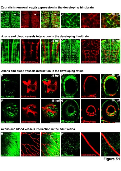
Neurons expressing VEGF-A are closely associated with blood vessels (related to Figure 1) (A) Confocal section of double in situ/immunolabelling with vegfaa and EGFP in the hindbrain of Tg(huc:egfp) line at 48 hpf shows neuronal expression of vegfa in the zebrafish brain. Close up of vegfaa expression in the hindbrain (A'). (B, C) Confocal section of double immunolabelling showing the physical interaction/close proximity between axons (Acetyl Tubulin, green) and blood vessels (red) in Tg(kdrl:mCherry) hindbrain at 36 and 48 hpf. (D-H) Confocal section of double immunolabelling showing the physical interaction/close proximity between axons (Acetyl Tubulin, green) and blood vessels (red) in Tg(kdrl:mCherry) retina at 36 hpf, 48 hpf or in the adult retina. Close up on a blood vessel in the adult retina (H'). Arrowheads show co-localization. Dorsal view of the brain with anterior up. Lateral view of the retina. Scale bars: 100 μm (A and D-H) or 10 μm (A', B, C and H'). |