
Fig. S1
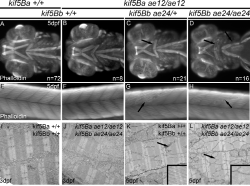
Muscle phenotypes of kif5Blof mutants. A-D) Ventral view of the anterior region of zebrafish at 5 dpf. Muscle fibers are shorter than Wt, and broken muscle fibers are detected along the body axis (arrows in C and D). This phenotype was not observed in kif5Ba single mutants (B). E-H) Lateral views of the tail at 5 dpf; broken muscle fibers are observed in kif5Blof mutants (arrows in G and H). I-L) Electron microscopy of the ocular muscle at 3 dpf (I, J) and 5 dpf (K, L). No disruption of muscle ultrastructure was apparent at 3 dpf (I, J). However, at 5 dpf the M-line of the sarcomere is diminished in kif5Blof mutants (I, J and insets). |
| Fish: | |
|---|---|
| Observed In: | |
| Stage: | Day 5 |