
Fig. S1
- ID
- ZDB-FIG-170907-19
- Publication
- Pfefferli et al., 2017 - The careg element reveals a common regulation of regeneration in the zebrafish myocardium and fin
- Other Figures
- All Figure Page
- Back to All Figure Page
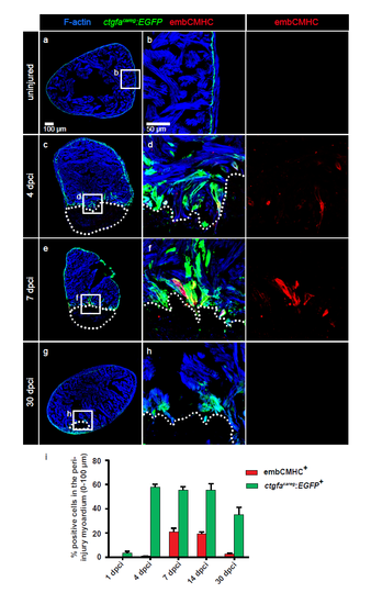
The ctgfacareg:EGFP transgenic reporter is transiently activated in the peri-injured myocardium during heart regeneration. (a-h) Immunofluorescence staining of ctgfacareg:EGFP transversal heart sections with antibodies against GFP (green) and embCMHC (red) at different time points after cryoinjury. The intact myocardium is detected by F-actin staining (Phalloidin, blue). (a-b) In uninjured ventricle, ctgfacareg:EGFP is expressed in a fine layer in the subcortical myocardium. No embCMHC is observed. (c-d) At 4 dpci, ctgfacareg:EGFP is induced in trabecular fascicles abutting the post-infarcted area, a subset of which also displays embCMHC expression. (e-f) At 7 dpci, ctgfacareg:EGFP expression is maintained at the injury border covering embCMHC+ CMs. (g-h) At 30 dpci, ctgfacareg:EGFP is reduced to a small margin around the remaining fibrotic tissue. (i) Quantification of ctgfacareg:EGFP+ and embCMHC+ area within 100 μm from the injury border. N ≥ 8. Post-infarcted ventricle is encircled with a dotted line. The same rules apply to all subsequent figures. |