
Fig. 2
- ID
- ZDB-FIG-170825-33
- Publication
- Wehner et al., 2017 - Wnt signaling controls pro-regenerative Collagen XII in functional spinal cord regeneration in zebrafish
- Other Figures
- All Figure Page
- Back to All Figure Page
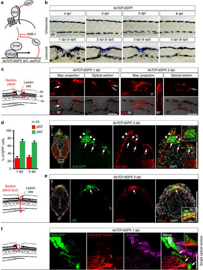
Wnt/β-catenin signaling is mainly active in fibroblast-like cells after spinal cord lesion. a Cartoon showing relevant transcriptional read-outs of Wnt/β-catenin pathway activity, feedback regulatory mechanisms and strategies to interfere with signaling at different levels of the pathway. Abbreviation: β-cat., β-catenin. b Detection of gfp mRNA in the 6xTCF:dGFP transgenic reporter line shows transient activity of the Wnt/β-catenin pathway in the lesion site during regeneration. c gfp expression is largely confined to the lesion edge in 6xTCF:dGFP transgenic larvae (arrow). A few additional cells are labelled within the presumptive spinal cord region (empty arrows). Abbreviations: nc, notochord; sc, spinal cord; fin, dorsal fin fold. d The 6xTCF:dGFP Wnt reporter is mostly active in subepidermal p63− cells (arrows; ≥ 69% of all 6xTCF:dGFP+ cells at 1 dpl and 2 dpl), but also in p63+ basal keratinocytes (arrowheads) in the lesion site. # indicates non-specific signal. e Consistent with a fibroblast identity, subepidermal cells (below dashed line) co-express col1a2 (red) and gfp mRNA (green) in a lesioned 6xTCF:dGFP transgenic animal (arrow). Dashed line indicates junction between basal epithelium and adjacent connective tissue. f Fibroblast-like (6xTCF:dGFP+/p63−) cells are found in close proximity to regenerating axons (anti-acetylated Tubulin+) in the spinal lesion site at 1 dpl (arrows). A single optical section is shown. a–f Views are lateral (b,c,f; dorsal is up, rostral is left) or transversal (d,e; dorsal is up) as indicated. BF: brightfield. Scale bars: whole mounts, 100 µm b,c and 25 µm f; sections: 100 µm. Error bars indicate s.e.m |
| Gene: | |
|---|---|
| Antibodies: | |
| Fish: | |
| Condition: | |
| Anatomical Terms: | |
| Stage Range: | Day 4 to Days 7-13 |