
FIGURE
Fig. 1
Fig. 1
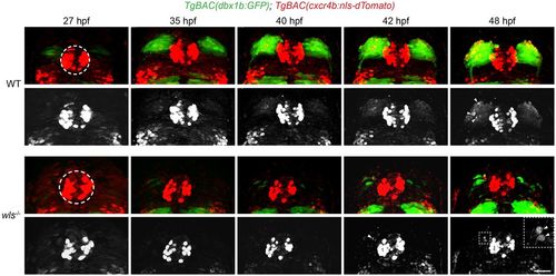
Live imaging of dbx1b+ and cxcr4b+ populations. Live imaging of WT and wls mutant TgBAC(dbx1b:GFP) and TgBAC(cxcr4b:nls-dTomato) double-labeled embryos between 27 and 48 hpf (Movie 1). Black and white images are of dTomato-labeled nuclei alone. dTomato-labeled nuclei are found in the pineal anlage (dashed circle) and, over time, in cells arising within the developing dHb (arrowheads and inset). Scale bar: 30 µm (11 µm for inset). |
Expression Data
| Genes: | |
|---|---|
| Fish: | |
| Anatomical Terms: | |
| Stage Range: | Prim-5 to Long-pec |
Expression Detail
Antibody Labeling
Phenotype Data
| Fish: | |
|---|---|
| Observed In: | |
| Stage Range: | Prim-5 to Long-pec |
Phenotype Detail
Acknowledgments
This image is the copyrighted work of the attributed author or publisher, and
ZFIN has permission only to display this image to its users.
Additional permissions should be obtained from the applicable author or publisher of the image.
Full text @ Development