
Fig. S4
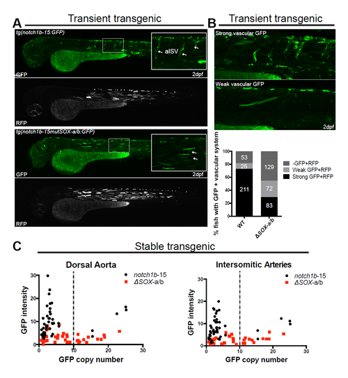
Sox sites ablation compromises notch1b-15 enhancer activity. (A) A representative confocal projection of wild-type zebrafish F0 larvae injected with notch1b-15:GFP (top panel) and notch1b-15:mutSOX-a/b:GFP (bottom panel). Bright GFP expression could be observed in the intersomittic vessel and dorsal aorta of fish injected with the wild-type notch1b-15 construct, whereas expression of GFP in fish injected with the mutant construct is almost completely absent from the arterial vascular network (white arrows). (B) Graph indicating levels of GFP expression in F0 zebrafish transgenic for notch1b-15:GFP and notch1b-15mutSOX-a/b:GFP transgenes. All scored fish were also positive for the internal control cardiac actin:RFP (+RFP). Number of zebrafish examined for each conditions was indicated, expression was scored as no (-), weak or strong GFP . (C) Scatter plots representing the relationship between genomic GFP copy number and GFP intensity in endothelial lining of dorsal aorta (left panel) and intersomittic vessels (right panel) in stable tg(notch1b-15) and tg(notch1b-15mutSOX-a/b:GFP). Only larvae with GFP copy number <10 were included in the analysis for Fig. 7B-C. Scored tg(notch1b-15), n=46; tg(notch1b-15mutSOX-a/b:GFP), n=42; larvae were each pooled from 4 independent founders. |