
Fig. S2
- ID
- ZDB-FIG-170703-10
- Publication
- McArthur et al., 2017 - Key Features of Structural and Functional Organization of Zebrafish Facial Motor Neurons Are Resilient to Disruption of Neuronal Migration
- Other Figures
- All Figure Page
- Back to All Figure Page
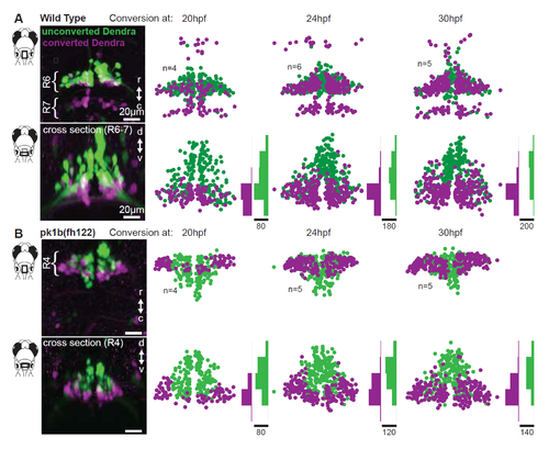
Facial motor neuron age topography in wild type and pk1b(fh122) mutants. Related to Figure 3. Zebrafish embryos expressed the photo convertible protein Dendra in cranial motor neurons and were exposed to ultraviolet light at three time points during facial motor neurons differentiation (20, 24 and 30 hpf), then imaged at 3 dpf. For each conversion time, neurons older than that time point contain converted (magenta/purple) Dendra, while younger neurons contain only unconverted (green) Dendra. (A) In wild type larvae, older facial motor neurons are located in the ventral-most part of the facial motor nucleus. (B) In pk1b(fh122) mutant larvae, facial motor neurons adopt the same dorsoventral age topography observed in wild type larvae. Note that there is a modest shift in the earliest conversion time point, such that the oldest mutant neurons are found laterally as well as ventrally. |