
FIGURE
Fig. 3
- ID
- ZDB-FIG-170608-12
- Publication
- Kawahara et al., 2016 - Characterization of Zebrafish Models of Marinesco-Sjögren Syndrome
- Other Figures
- All Figure Page
- Back to All Figure Page
Fig. 3
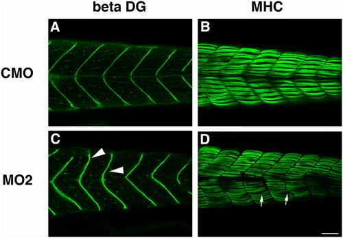
Immunohistochemistry of skeletal muscle tissue of morpholino-injected fish. Immunostaining of CMO injected fish (A, B) and sil1 morphant (MO2) injected fish (C, D) with antibodies against beta-dystroglycan (beta-DG) (A, C) and myosin heavy chain (MHC) (B, D). Beta-dystroglycan expression at the myosepta of MO2 injected 4 dpf embryos is misshapen and has a less clear v-shaped structure. Staining with anti-MHC indicated that formation of myofibers is disturbed in MO2. Arrowheads indicate the disturbance of myosepta in MO2-injected fish. Bar: 100 μm. Arrows indicate the abnormal structure of myofibers. |
Expression Data
| Gene: | |
|---|---|
| Antibodies: | |
| Fish: | |
| Knockdown Reagent: | |
| Anatomical Terms: | |
| Stage: | Day 4 |
Expression Detail
Antibody Labeling
Phenotype Data
| Fish: | |
|---|---|
| Knockdown Reagent: | |
| Observed In: | |
| Stage: | Day 4 |
Phenotype Detail
Acknowledgments
This image is the copyrighted work of the attributed author or publisher, and
ZFIN has permission only to display this image to its users.
Additional permissions should be obtained from the applicable author or publisher of the image.
Full text @ PLoS One