
Fig. 4
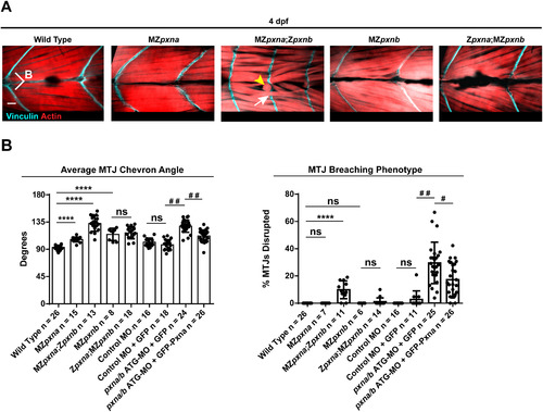
Paxillin mutants exhibit defects in myotome development. (A) Vinculin and Actin were visualized in 4 dpf embryos to show MTJs and myofibers respectively. Chevron angle (B) was measured for three consecutive MTJs and averaged for each embryo. MZpxna;Zpxnb embryos exhibited over-elongated myofibers passing through MTJs (white arrow) in some of their myotomes. Myofiber detachment and retraction (yellow arrowhead) was also observed. Zpxna;MZpxnb embryos had more mild MTJ breaching defects at this stage. Scale bar=10 µm (B) MTJ chevron angles and disrupted MTJs were quantified at 4 dpf in wild-type embryos, Paxillin MZ single mutants, Paxillin MZ double mutants and wild-type embryos injected with either control MO or pxna/b ATG-MO. Ectopic expression of GFP-Pxna fusion protein partially rescued myotome defects in pxna/b ATG-MO injected embryos. Data points represent individual embryo means and error bars show standard deviations from three independent experiments. **** p<0.001 as determined by ANOVA/Fisher's LSD post-hoc test, # # p<0.01 as determined by T-test, ns=not significant. |
| Antibodies: | |
|---|---|
| Fish: | |
| Anatomical Terms: | |
| Stage: | Day 4 |
| Fish: | |
|---|---|
| Knockdown Reagent: | |
| Observed In: | |
| Stage: | Day 4 |
Reprinted from Developmental Biology, 425(1), Jacob, A.E., Amack, J.D., Turner, C.E., Paxillin Genes and Actomyosin Contractility Regulate Myotome Morphogenesis in Zebrafish, 70-84, Copyright (2017) with permission from Elsevier. Full text @ Dev. Biol.