
Fig. S9
- ID
- ZDB-FIG-170508-4
- Publication
- Armstrong et al., 2017 - Shh promotes direct interactions between epidermal cells and osteoblast progenitors to shape regenerated zebrafish bone
- Other Figures
- All Figure Page
- Back to All Figure Page
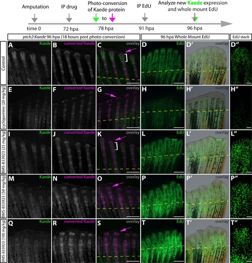
The smoothened inhibitor BMS-833923 does not exhibit the global antiproliferative effect of cyclopamine on regenerating zebrafish fins. (A-T) Whole-mount fin images showing ptch2:Kaede expression and EdU incorporation in DMSO (A-D''), cyclopamine (20 mg/kg) (E-H''), BMS-833923 (25 mg/kg) (I-L''), BMS-833923 (50 mg/kg) (M-P''), or BMS- 833923 (100 mg/kg) (Q-T'') treated fish at 96 hpa. At 72 hpa, each fish was injected intraperitoneally with the indicated small molecule or DMSO (control). Kaede protein was photoconverted at 78 hpa and tissue was collected at 96 hpa after a 5 hour pulse with EdU. New Kaede protein is shown in green and photoconverted Kaede in magenta. At least four fish were analyzed from each group. The white bracket indicates the domain of new Kaede expression since photoconversion and the magenta arrows denote previously Hedgehog/Smoothened responsive epidermis. EdU-incorporating nuclei are green in panels D, H, L, P, and T. Overlays with corresponding brightfield images are shown (D', H', L', P', T'). Panels D'', H'', L'', P'', T'' show confocal images of individual rays from each sample. Note: control (A-D), cyclopamine (20 mg/kg) (E-H), and BMS-833923 (50 mg/kg) (M-O) images are also shown in Figure 5. Scale bars for D'', H'', L'', P'', T'': 50 μm. All other scale bars: 500 μm. |