|
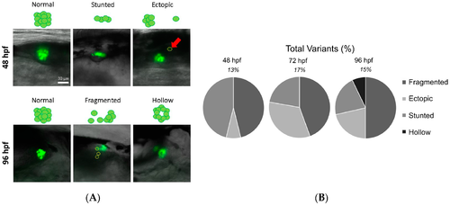
Islet variant morphologies observed within DMSO-treated embryos change throughout development. To complement the distributions of islet areas, islet morphologies were examined for morphological variants at 48, 72, 96, and 168 hpf. (A) Representative images of islet variants at 48 and 96 hpf are shown; (B) The prevalence of these variants changes over time; percentages shown are the total prevalence of variants among all DMSO-treated embryos. Pie charts show the distributions of each of the variant morphologies within the total number of variants at each time point. At 168 hpf, only one fragmented islet was observed; therefore, data variation could not be shown. All images are of the right lateral side, acquired with a 20 × objective, shown in a posterior (left) to anterior (right) orientation.
|