
Fig. S3
- ID
- ZDB-FIG-170321-20
- Publication
- Zhang et al., 2017 - Left Habenula Mediates Light-Preference Behavior in Zebrafish via an Asymmetrical Visual Pathway
- Other Figures
- All Figure Page
- Back to All Figure Page
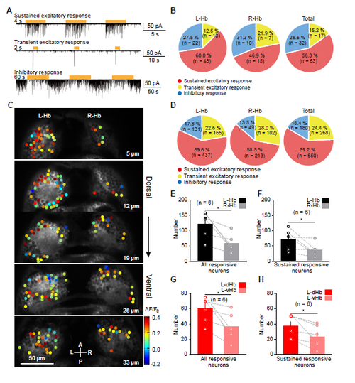
Physiological Characterization and Spatial Distribution of Visually Responsive Hb Neurons by Using In Vivo Whole-cell Recording and Confocal Calcium Imaging. (A) Example traces showing that visually responsive Hb neurons can be classified into three types, which exhibits sustained excitatory, transient excitatory, or inhibitory responses, respectively. The cells were held at -60 mV. The brown thick lines represent flash stimuli. (B) Percentages of the three types of visually responsive neurons in the L-Hb, R-Hb, and whole Hb. Data were obtained through in vivo whole-cell recording. (C) Calcium activities of neurons in all the five Hb layers (5μm, 12μm, 19μm, 26μm, and 33 μm away from the dorsal) in a 6-dpf Tg(HuC:GCaMP5.0)ion13dTg larva in response to 10-s flash stimuli. The response amplitude is color-coded. A, anterior; L, left; P, posterior; R, right. (D) Percentages of the three types of visually responsive neurons in the L-Hb, R-Hb, and whole Hb. Data were obtained through in vivo calcium imaging. (E and F) Mean numbers of all responsive (E) or sustained excitatory responsive (F) neurons in the L-Hb and R-Hb for each larva. (G and H) Mean numbers of all responsive (G) or sustained excitatory responsive (H) neurons in the L-dHb and ventral L-Hb (L-vHb) (the first two dorsal layers versus the last two ventral layers) for each larva. The data used in (D - H) are the same with those in Figures 4G and 4H. The numbers in the brackets represent the numbers of neurons (B and D) or larvae (E-H) examined. Error bars, SEM. *p < 0.05 (paired two-tailed Student’s t test). |