
Fig. 3
- ID
- ZDB-FIG-170314-6
- Publication
- Williams et al., 2017 - Cyp1b1 Regulates Ocular Fissure Closure Through a Retinoic Acid-Independent Pathway
- Other Figures
- All Figure Page
- Back to All Figure Page
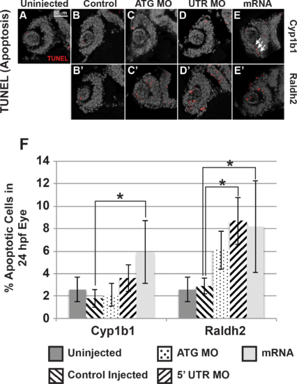
Cyp1b1 and RA regulated cell survival in the developing eye. Cyp1b1 ATG (C) and 5′ UTR (E) MO knockdown did not significantly affect apoptosis in the eye at 24 hpf compared to uninjected (A) and control-injected (B) embryos. Cyp1b1 overexpression (E) increased apoptosis and the apoptotic cells were specifically localized to the inferior ocular fissure (arrows). Raldh2 5′ UTR (D') but not ATG (C') MO knockdown significantly decreased cell survival throughout the developing eye compared to uninjected (A) and control injected (B'). Similarly, overexpression of raldh2 (E') increased apoptosis in the eye. The percentage of apoptotic cells in 24 hpf developing eyes (F) of embryos injected with Cyp1b1 MO (ATG or 5′ UTR), cyp1b1 mRNA, Raldh2 (ATG or 5′ UTR) MO, raldh2 mRNA compared to uninjected and control injected (*P < 0.01). |
| Fish: | |
|---|---|
| Knockdown Reagent: | |
| Observed In: | |
| Stage: | Prim-5 |