
Fig. S7
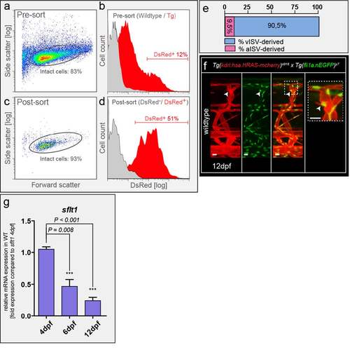
Spinal cord vascularization in WT zebrafish (a,b) Neuronal cells were isolated from Tg(Xla.Tubb:DsRed)zf148 embryos at 3dpf by FACS with indicated gating settings. About 12% of all intact cells were DsRed+ neurons prior to sorting (Pre-sort). (c,d) Post-sorting analysis showed that sorted neuronal cells are enriched to 51% neuronal DsRed+ cells. DsRed- cells contained less than 1.7 % DsRed+ cells. (e) Percentage of aISV and vISV giving rise to sprouts at level of neural tube in late stage WT embryo; 400 ISVs in n=20 embryos. (f) Nuclear positioning in sprout contributing to spinal cord vascularization in wildtype Tg(kdrl:has.HRAS-mcherry)s916;Tg(fli1a:nEGFP)y7 at 12dpf; representative image from 6 embryos (g) Quantitative PCR for sflt1 at indicated time points. Note decreased expression of sflt1 associates with sprout appearance and spinal cord vascularization. mean ± s.e.m, n=3 experiments, 30 embryos/experiments dpf, days post fertilization; ISV, intersegmental vessel (a – artery, v – vein). Scale bar, 10μm in f. |