|
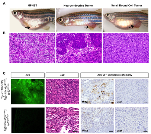
MPNSTs arising in Tg(mitfa:atg5K130R); tp53M214K/+ fish express sox10. A.-B. Representative photomicrographs of gross macroscopic appearance of tumors A. and hematoxylin and eosin (H&E) stained microscopic sections of corresponding tumors B. in Tg(mitfa:atg5K130R); tp53M214K/+ fish. MPNST, malignant peripheral nerve sheath tumor. Scale bars, 50 μm. See Table 1 for details of numbers of fish with each tumor type. C. Representative confocal images to detect GFP expression, H&E staining, and anti-GFP immunoperoxidase staining of an MPNST from a Tg(mitfa:atg5K130R); Tg(sox10:EGFP); tp53M214K/+ fish (top) and an MPNST from a GFP-negative control Tg(mitfa:atg5K130R); tp53M214K/+ (bottom). Liver is shown for the anti-GFP immunoperoxidase staining as a representative non-tumor tissue (right panels). Scale bars, 100 µm.
|